Author: Dr. Astrid Bernaga.
In our days, we live in a technologic era with the benefit of digital simulation tools. This technology can help us to learn the morphology of our bodies, but the corpse experience for the medical students is crucial for studying anatomy and to have respect for the human life.
Using cadavers for dissection is a tradition since the 16th century and it is the gold standard for teaching anatomy. It is the best method to practice open and minimally invasive procedures for students of bioengineering, medicine students, and different surgical specialties (trauma, general surgery, neurosurgery and gastrointestinal) are using this method for training. Indeed, this traditional practice for so many years is evident because the corpses are high fidelity models and the variability between them is fundamental to learn Anatomy (Yiasemidou, Marina; Gkaragkani, E; Glassman, D; Biyani, C.S; 2017).
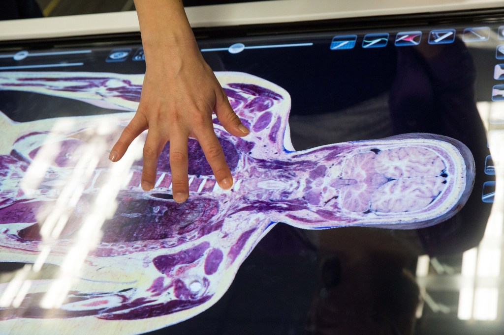
Initial approaches to “virtual anatomy” emerged in the 1990s, still a long way to go. A crucial development in this context was the Visible Human Project of the National Library of Medicine (NLM). Two human donors were frozen and cut into thin slices, which were then photographed and digitalized (Fellner, Engel, & Kremer, 2017). Anatomage is useful in this case, because of the cross-sections images of the complete human body, help to train radiology students in 3D and 2D anatomy, like the Computerized Tomography (CT) and Magnetic Resonance Imaging (MRI) (Ward, Wertz, & Mickelsen, 2018). Virtual Reality (VR), and Augmented Reality are valuable technologies for teaching anatomy, but students can suffer adverse effects, such as headaches (25%), dizziness (40%) and blurred vision (35%) (Moro, Štromberga, Raikos, & Stirling, 2017). 3D printing could be the most useful tool, but only for the osteopathic system. The 3D printing could easily simulate the bones, the rest of the tissues still cannot be imitated (Yousef, 2015).
The emotions and values are essential in the formation of a future physician. In fact, cadaveric dissection induces both positive and negative experience in students (Lima, Tinoco, Coelho-Ferraz, De Sousa, & Francesquini Júnior, 2017). In Kaundal & Kaundal, 2017, before the examination, the students feel fear (93%), excitement (68%), interest (64%) and depression (25%) in that order. But after starting dissection, their emotions change to excitement (91%), interest (89%), fear (15%) and depression (7%). Regardless of the symptoms experienced by the students, 94% of them were determined, interested and ready to study anatomy with cadaveric dissection. Most of all, the students considered this examination the best tool to study anatomy and had shown a sense of gratitude to the people and the relatives who had donated their bodies.
Currently, there is an unavailability of corpses worldwide for causes like religion, culture and more medical schools. This means that thousands of medical graduates had never practice on a real body (Morán, 2013). Most importantly, this can result in severe errors that end in thousands of avoidable deaths per year on the operating room tables. Also, it has been reported an increase of confidence in the Operating Room (OR). This confidence is maintained up to seven months after the training sessions (Yiasemidou, Marina; Gkaragkani, E; Glassman, D; Biyani, C.S; 2017).
Not many medical schools have a program to donate your body after death. But in Stanford Medicine and UCSF (University of California San Francisco), you can help science if you give your body. Both programs to become a donor are called “Willed Body Program.” Your body is going to be respected, and after that, returned your ashes to your relatives. For more info visit Stanford Medicine http://med.stanford.edu/anatomy/donate.html and UCSF http://willedbodyprogram.ucsf.edu
Finally, knowing these technologies, we might think is the future of teaching. However, the cost of technologies for schools in developing countries, the low accuracy and their lack of developing deep emotions creates an environment of a coexistence more than for a replacement. Cadaveric dissection is the gold standard tool for anatomy study for the training of physicians, who need to be close to death in the early stages of their formation. In other words, understanding death and human life.
And… Would you donate your body to science?
______________________________________________________
Bibliography
Fellner, F., Engel, K., & Kremer, C. (2017, July 27). Virtual Anatomy: The Dissecting Theatre of the Future- Implementation of Cinematic Rendering in a Large 8K High-Resolution Projection Environment. J. Biomedical Science and Engineering, pp. Vol. 10 (No.8) 367-375.
Kaundal, A., & Kaundal, e. a. (2017, Aug 5). Perception of students towards cadaveric dissection in a tertiary case teaching institution. Int J Res Med Sci, pp. (8) 3684-3687.
Lima, L., Tinoco, R., Coelho-Ferraz, M., De Sousa, E. D., & Francesquini Júnior, L. &. (2017, June). Respect for the Donated Corpse in the View of Dentistry and Medicine Students. Int. J. Ortodontostomat, pp. 11(2) 141-146.
Morán, C. (2013, January 22). Ser médico sin tocar un cadáver. Retrieved from El País: https://elpais.com/sociedad/2013/01/21/actualidad/1358788802_664958.html
Moro, C., Štromberga, Z., Raikos, A., & Stirling, A. (2017, April 17). The effectiveness of virtual and augmented reality in health sciences and medical anatomy. Anat Sci Educ, pp. (10) 549-559.
Stanford Medicine. Retrieved from Willed Body Program: http://med.stanford.edu/anatomy/donate.html
University of California San Francisco (UCSF). Retrieved from Willed Body Program: http://willedbodyprogram.ucsf.edu/
Ward, T. M., Wertz, C. I., & Mickelsen, W. (2018, January/February Vol. 89). Anatomage Table Enhances Radiologic Technology Education. Radiologic Technology, pp. (3) 304-306.
Yiasemidou, Marina; Gkaragkani, E; Glassman, D; Biyani, C.S; (2017). Cadaveric simulation: a review of reviews. Ir J Med Sci, https://doi.org/10.1007/s11845-017-1704-y
Yousef, A. e. (2015). The application of 3D printing in anatomy education. Retrieved from Medical Education Online: http://dx.doi.org/10.302/meo.v20.29847
Top photo by Deanna Dent/ASU

Leave a comment